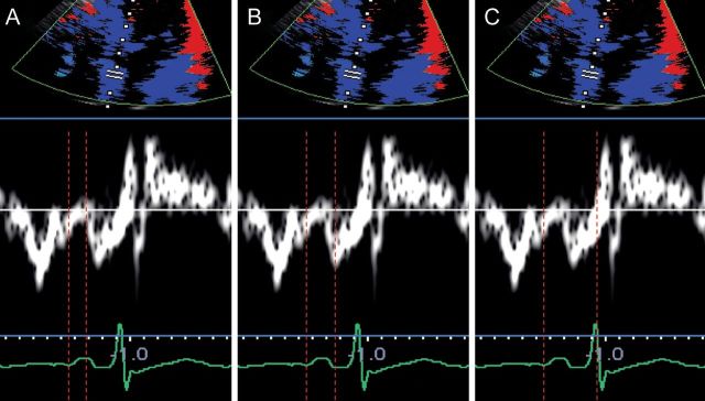
Figure 3.

Left atrial electromechanical intervals measured through tissue Doppler imaging: p-A start interval, measured from the onset of the P-wave to the onset of A′ (A); p-Apeak interval, measured from the onset of the P-wave to the peak of A′ (B); total electromechanical time, measured from the onset of the P-wave to the end of A′ (C).
