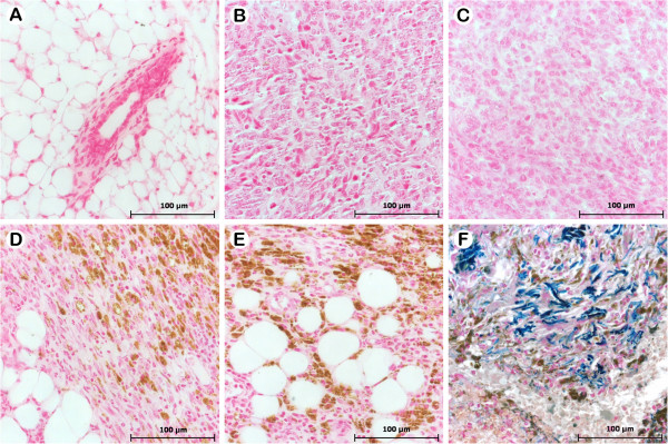
Figure 6.
Histopathology of mammary gland and tumors developed from Balb/c mice bearing 4T1 breast carcinoma in different treatment groups. The animals were treated as described in section 2.3 and tumor slides were stained with Perls coloration. A) Breast tissue of healthy group. B) 4T1 tumor cells without treatment (control), with treatment of Rh2(H2cit)4 (C), Magh-Rh2(H2cit)4 (D) and Magh-citrate (E). F) Positive control staining Perls Prussian blue (human tissue). The blue-green indicates Fe2+ (iron is non-oxidized) and brown indicates the presence of Fe3+ (oxidized iron). The presence of maghemite nanoparticles (oxidized iron) was observed, represented by brown coloration.

