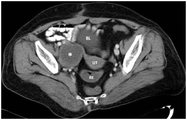
Figure 1.
Axial, enhanced computed tomography image of the pelvis demonstrates a mass in the anatomic region of the right ovary, corresponding to the ovary’s schwannoma.
Note: The enlarged ovary is well delineated, with internal low density and enhanced peripherally.
Abbreviations: UT, uterus; BL, urinary bladder; RE, rectum.

