
Figure 9.
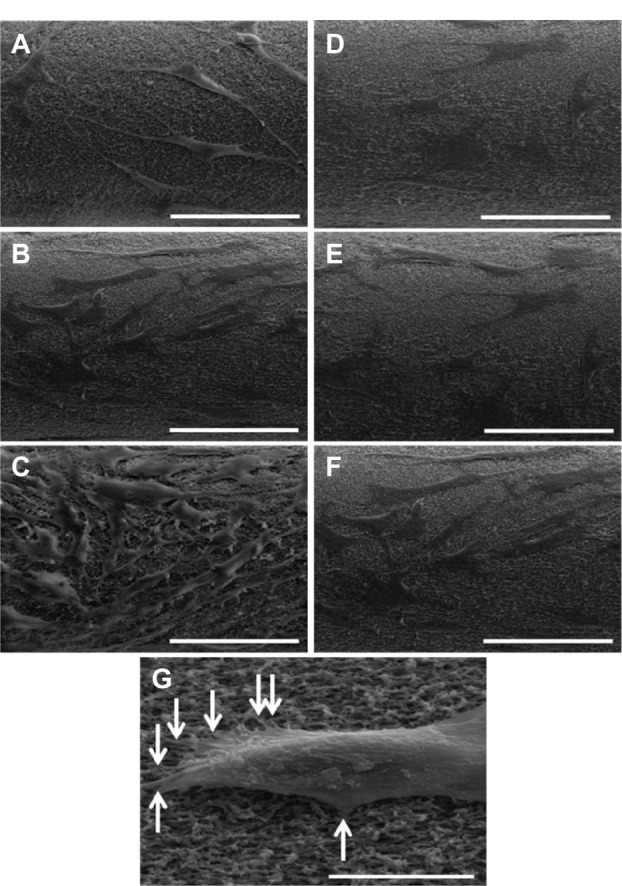
Representative scanning electron microscopy images of cell attachment on Fbg fibers. Images A, B, and C are FN-grafted Fbg fibers showing fibroblast cell attachment and proliferation after 24 hours, 3 days, and 5 days, respectively. Images D, E, and F are bare Fbg fibers after 24 hours, 3 days, and 5 days of fibroblast proliferation, respectively, (scale bar: 100 μm). Image G shows single-cell attachment with fiber pore structures (denoted by white arrows; Scale bar: 50 μm).