Figure 1.
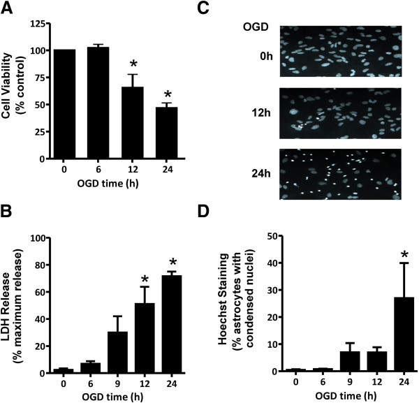
OGD induces both necrotic and apoptotic cell death in cultured cortical astrocytes. A) Cell viability of cultured cortical astrocytes, as assessed by the metabolic MTT assay following 0-24 h OGD followed by 24 h reperfusion. B) Necrotic cell death measured by lactate dehydrogenase (LDH) assay following 0-24 h OGD followed by 24 h reperfusion. C) Representative photomicrographs of primary CD1 cortical astrocytes cultures that were OGD treated for 0, 12, and 24 hours followed by 24 h reperfusion and stained with Hoescht dye to reveal cells dying by apoptosis as evidenced by condensed chromatin/ pyknotic nuclei. D) Quantification of apoptotic CD1 astrocytic cell death following 0-24 h OGD followed by 24 h reperfusion. All data represents the mean ± SD of 5 independent experiments and were analyzed by ANOVA followed by Dunnet’s post-hoc testing, * p<0.05 versus untreated control cultures.

