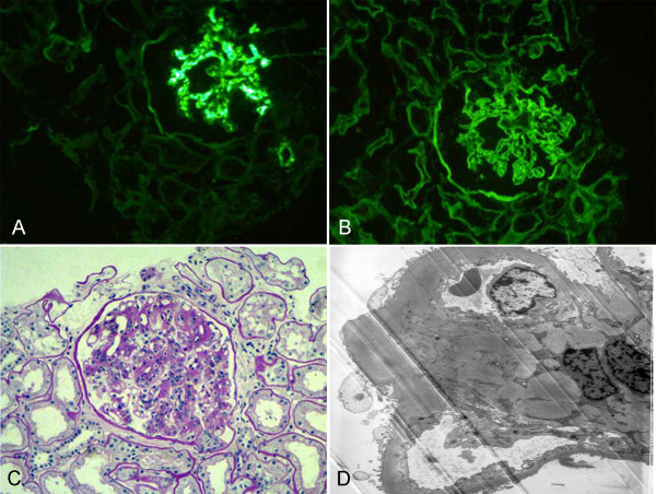
Figure 1.
A case with immunoglobulin A (IgA) nephropathy and diabetic nephropathy (DN). (A) Typical mesangial absorbance pattern after labeling with anti-IgA antibody (IF, 200×). (B) The deposits of mainly IgG collected in the basement membrane and appeared in the linear pattern as shown by immunofluorescence (IF, 200×). (C) Mesangial cellularity and matrix increased, and there was a thickening of the glomerular basement membrane (GBM) (PAS, 200×). (D) The electron micrograph demonstrated an increase in small dense deposits in the mesangium and the mesangial matrix. The basement membrane was diffusely thickened due to diabetic involvement (EM, 5000×). DN, diabetic nephropathy; EM, electron microscopy; GBM, glomerular basement membrane; IF, immunofluorescence; IgA, immunoglobulin A; IgG, immunoglobulin G; PAS, periodic acid-Schiff.

