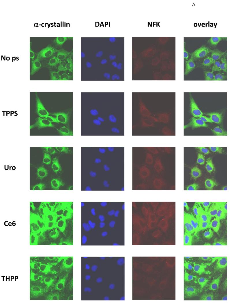
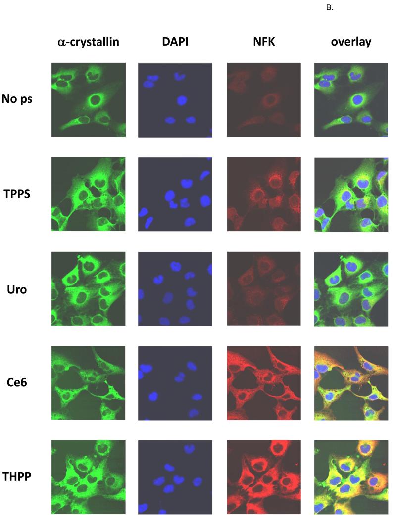
Figure 7.
Confocal fluorescent microscopy of (A) dark-incubated and (B) UVA-irradiated HLE cells. Nearly confluent HLE-B3 cells were treated overnight with or without 10 μM porphyrins as indicated and after washing were kept in the dark. Fixed cells were simultaneously stained with rabbit anti-NFK and mouse anti-αA and anti-αB-crystallin followed by staining with Alexa Fluor anti-rabbit 568 and anti-mouse 488 and then with DAPI. α-crystallin (green), DAPI (blue), NFK (red), and overlay of all three.


