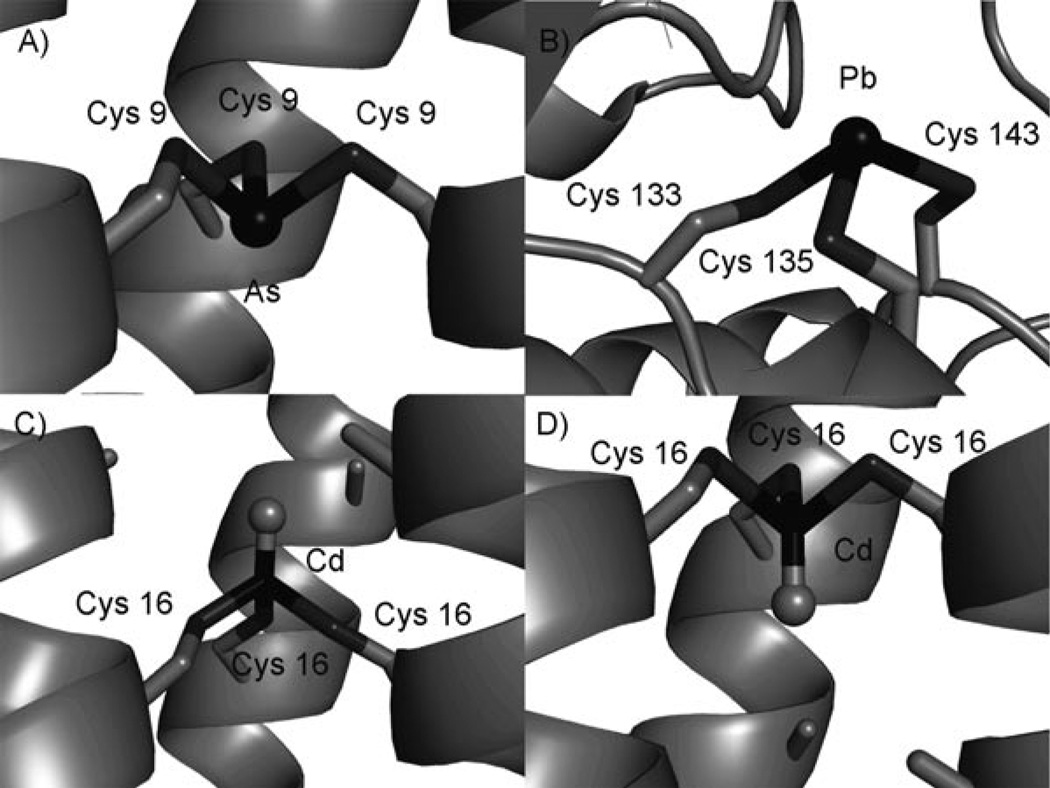
Figure 5.
a) Side-view of AsIII coordinated to the Cys residues in CSL9C.[50] b) Drawing generated using the X-ray crystal structure of PbII bound to ALAD (pdb: 1QNV).[56] Models, based on the X-ray crystal structure of As(CSL9C)3,[50] of CdS3O bound c) to TRIL12AL16C in the exo conformation and d) to TRIL16CL19A in the endo conformation. All figures were generated using PyMOL.

