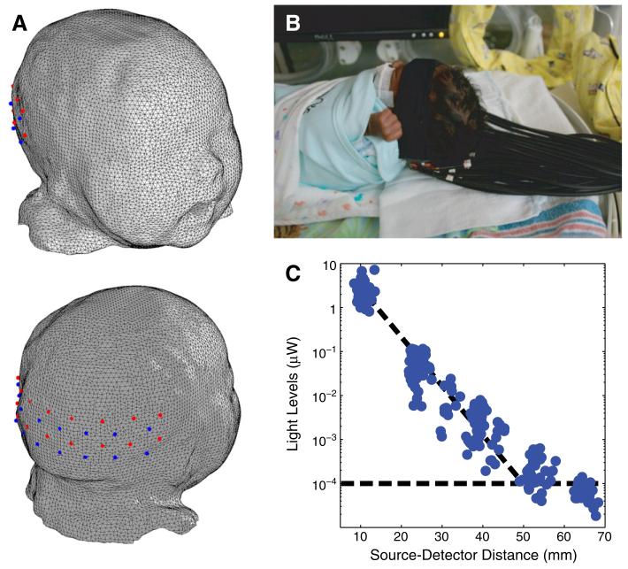
Fig. 1.
Bedside functional imaging of infants with DOT. (A) Infant head model and visual cortex imaging pad with 18 sources (red) and 16 detectors (blue), placed over the occipital cortex. (B) Photograph of the optical probe on a premature infant in an isolette. (C) Detected light level vs. source-detector separation on an infant. All first- and second-nearest neighbor pairs were detected simultaneously and were well above the noise floor (shown by the horizontal dotted line).

