
Figure 6.
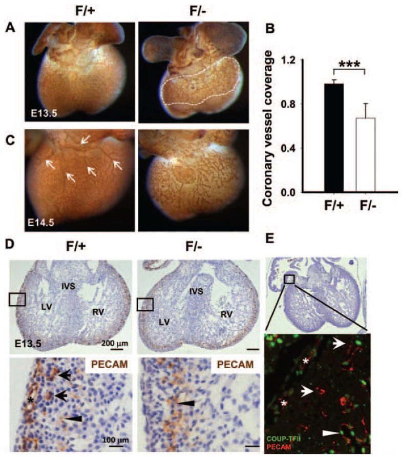
Inactivation of chicken ovalbumin upstream promoter-transcription factor II (COUP-TFII) leads to abnormal coronary angiogenesis. A and C, Whole-mount immunostaining of E13.5 and E14.5 mouse hearts with platelet-endothelial cell adhesion molecule (PECAM) illustrates vascular endothelium. A, A representative littermate control heart shows the normal vascular plexus in the ventricles. The vascular plexus of COUP-TFII mutant ventricle fails to extend to the apex of the heart. B, The coronary plexus coverage (ratio of region within white dotted line to total projected ventricular area) at E13.5 was significantly decreased in mutants. Error bars indicate SD; **P<0.01. C, A vessel with 4 major branches (arrows) forms on the dorsal side of the control ventricle, whereas COUP-TFIIF/− mutant ventricles at E14.5 present abnormal coronary morphogenesis, with less extensive vascular network and defective branching. D, Histological sections of PECAM-stained control and mutant hearts at E13.5 indicating that COUP-TFIIF/− mutants lack subepicardial vessels (asterisk) and intramyocardial vessels (arrows). Arrowheads denote endocardial PECAM staining. E, Hematoxylin and eosin and immunofluorescence stainings of wild-type mouse hearts at E13.5 reveal that COUP-TFII is expressed in subepicardial vessels (asterisks), intramyocardial vessels (arrows), and endocardium (arrowhead). LV indicates left ventricle; RV, right ventricle; IVS, interventricular septum.