Figure 7.

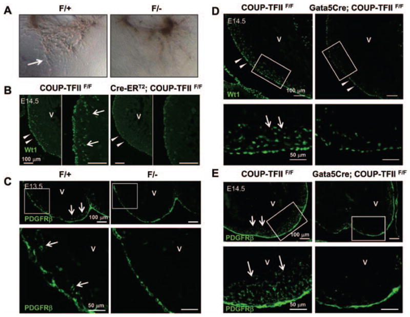
Epicardial-mesenchymal transformation (EMT) and epicardial migration are perturbed in chicken ovalbumin upstream promoter-transcription factor II (COUP-TFII) mutant hearts. A, Impaired EMT in COUP-TFIIF/− mutant epicardium. Representative figures of ex vivo heart explants on collagen gel. Cells migrate out of the control heart explants and transform to spindle-shaped mesenchymal cells (arrow), whereas little or no mesenchymal cells are observed in mutant heart explants. B and D, Decreased expression of Wilms tumor 1 (Wt1) in the epicardium of Cre-ERT2; COUP-TFIIF/F mutant hearts at E14.5 (with tamoxifen administration at E11.5) (B) or Gata5Cre; COUP-TFIIF/F mutant hearts at E14.5 (D) compared with littermate control COUP-TFIIF/F hearts. Numerous Wt1-positive epicardial derivatives were detected in the myocardium of controls (arrows), whereas decreased number of Wt1-positive epicardial derivatives was found in mutant hearts (white arrows). C and E, A decreased number of platelet-derived growth factor receptor β (PDGFRβ)–expressing epicardial-derived cells in the myocardium of COUP-TFIIF/− mutant hearts at E13.5 (C) or Gata5Cre; COUP-TFIIF/F mutant hearts at E14.5 (E) compared with littermate control hearts. Numerous PDGFRβ-positive epicardial derivatives were detected in the myocardium of controls (arrows), whereas a decreased number of PDGFRβ-positive epicardial derivatives were found in mutant hearts. V indicates ventricle.
