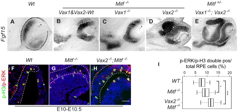
Figure 4. FGF-MAP kinase signaling regulates RPE-to-retina transition in Mitf mutants.
(A–E) In situ hybridization for Fgf15. In wild type (A), Fgf15 is normally restricted to the neural retina but is absent in the distal retina (arrow). (B–E) Ectopic expression of Fgf15 in the dorsal RPE is seen in Mit −/− (B), Vax2−/−;Mitf −/− (D), and Vax1−/−;Vax2−/−;Mitf +/− (E) though not in Vax1−/−;Mitf −/− mutants (C). Note that as in wild type, the dorsal future ciliary margin shows little Fgf15 labeling (arrow in B–E). (F–H) Increased numbers of p-H3/p-ERK double-positive cells in the RPE of E10−10.5 Mitf −/− (G) and Vax2−/−;Mitf −/− mutant (H) as compared to wild-type RPE (F). (I) Quantitation of the results obtained from sections as in F–G. Box plots show minimal, 25th percentile, median, 75th percentile and maximal percentages. Significance determined by Student’s t-test: *: p<0.05; ***: p<0.001 (2–3 sections per embryo, 6–10 embryos per genotype). Scale bar: 150 µm (A–E), 25 µm (F–H).

