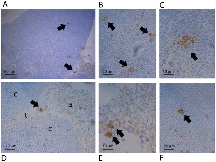
Figure 4. Identification of macrophages as the RRV-infected cells in the thymus, and their location.
Thymus sections from 8 infants of each mouse strain were examined at the peak of thymic infection by immunohistochemistry. A, Representative section showing infected macrophages in the cortex and adipose tissue (×100). B, Three infected macrophages in the cortex (×300). (C) Infected macrophage in the cortex (×600). (D) Higher magnification (×300) image of an infected macrophage from A in the adipose tissue (a) near the trabeculae (t) and cortex region (c). (E) Two infected macrophages in the adipose tissue under the thymus capsule (×600). F, Rare infected macrophage in the cortex region of the NOD thymus (×300). All images except F are from thymuses of BALB/c mice at 4 days post infection. No cells positive for RRV were seen in matching sections stained with the negative control antibody, or in thymus sections from mock-infected mice of the same strain at the same day post-infection.

