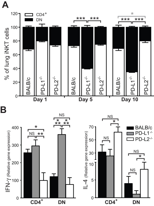
Figure 7. iNKT cell subpopulations are modulated in PD-L1−/− and PD-L2−/− animals after IAV infection.

(a) Wild type BALB/c, PD-L1−/− and PD-L2−/− mice (n = 3) were infected with IAV intranasally. Lungs were collected at day 1, 5 and 10 post infection and the frequency of CD4+ and DN iNKT cells determined by flow cytometry and compared (DN: black asterisk, CD4+: gray asterisk). (b) CD4+ and DN iNKT cells subsets were sorted from IAV infected lungs on day 5 from PD-L1−/−, PD-L2−/− and wild type mice BALB/c and IL-4 and IFN- γ were analysed by quantitative RT-PCR normalized to β-actin levels (n = 8). Data presented as mean ± SEM. * P>0.05, ** P>0.01 and *** P>0.001 as calculated by a one-way ANOVA with Tukey post hoc test and is representative of five independent experiments.
