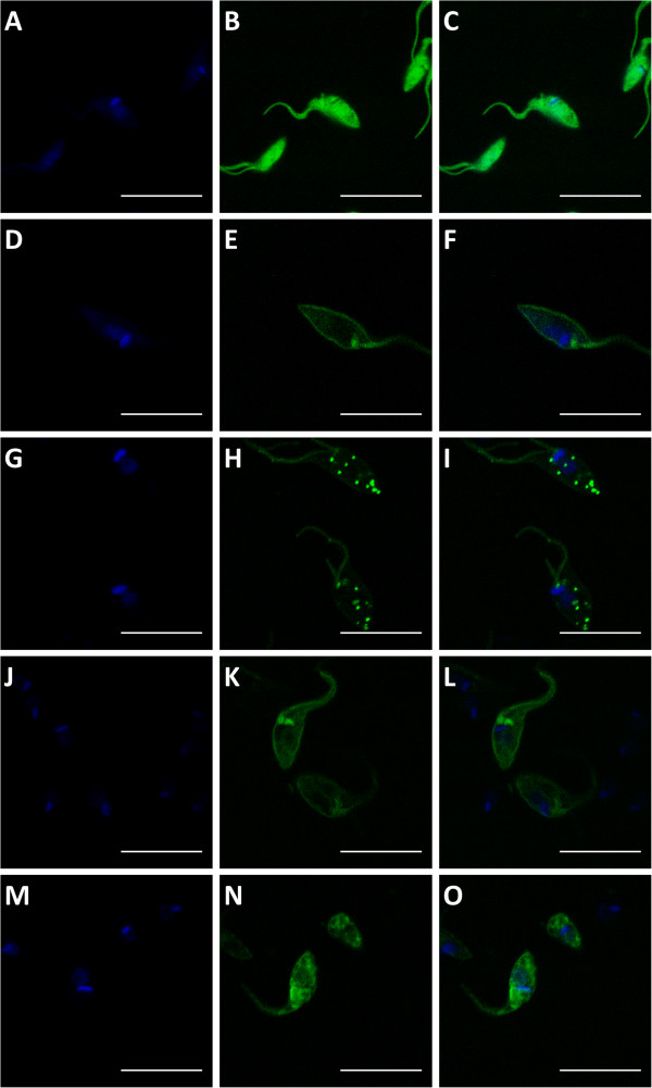
Figure 4.
Subcellular localization of distinct amastins in fusion with GFP. Images from stable transfected epimastigotes of the CL Brener or G strains obtained by confocal microscopy using 1000x magnification and 2.2 digital zoom. In panels (A-C), parasites transfected with a vector containing only GFP; (D-F), parasites transfected with δ-amastinGFP; (G-I), parasites transfected with δ-Ama40GFP; (J-L), parasites transfected β1-amastinGFP; (M-O), parasites transfected with β2-amastinGFP. DAPI staining are shown in panels (A, D, G, J and M); GFP fluorescence in panels (B, E, H, K and N) and merged images in panels (C, F, I, L and O). (Bar = 10 μm).

