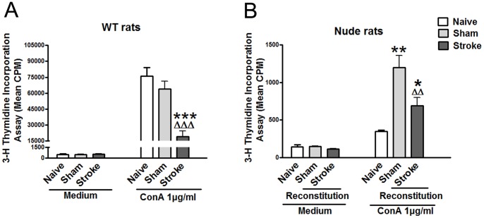
Figure 7. T cell proliferative assay after stroke.
Splenocytes were prepared 72 h after stroke from naïve, sham surgery and stroke animals. Cultured splenocytes were stimulated with 0 μg/ml, 1 μg/ml Con A. The cells were incubated for 66 h at 37°C and 5% CO2. [3H]-Thymidine was added to the culture and incubated for 18 h. Cells were harvested onto a glass fiber filter. The amount of radioactivity that integrated into divided lymphocytes was measured by a beta-plate scintillation reader. Results are expressed as counts per minute (cpm). A. Splenocytes proliferation in WT SD rats (n = 3–6/group). B. Splenocytes proliferation in T cells reconstituted nude rats (n = 3–5/group). Nude rats were reconstituted by intravenous injection of 1×10⁸ splenocytes in 100 μl RPMI 1640, and dMCAO was performed 24 h after reconstitution. *, **, *** vs. naïve, Δ, ΔΔ, ΔΔΔ vs. sham, p<0.05, 0.01, 0.001, respectively.

