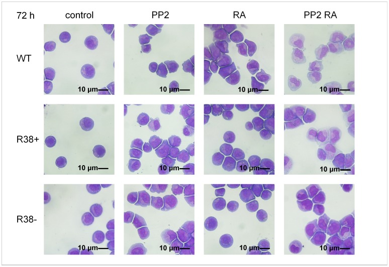
Figure 6. Wright’s stain cytology for control and treated WT, R38+ and R38− HL60 cell lines.
Control WT cells are round; RA-treated R38+ and R38− also retain a round, stem-like appearance. WT HL60 cells showed morphological changes consistent with differentiation toward granulocytes when treated with RA, PP2, or PP2+RA. Meanwhile the two RA-resistant HL60 cell lines, R38+ and R38−, both showed morphological changes consistent with differentiation only during PP2 and PP2+RA treatment, but not RA treatment alone.

