Figure 4.

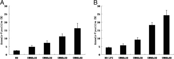
SWNHs promoted cell apoptosis of N9 cells, especially in pre-treated with LPS. After the cells had been cultured onto SWNHs-coated dishes for 48 h, the effect of SWNHs on cell apoptosis distribution was determined by flow cytometry. Apoptosis of N9 cells (A) and N9 cells pre-treated with LPS (B) was promoted with the increasing concentrations of SWNHs (P < 0.001). The effect on apoptosis induced by SWNHs on N9 cells pre-treated with LPS was more significant than N9 cells. All data are represented as mean ± SEM.
