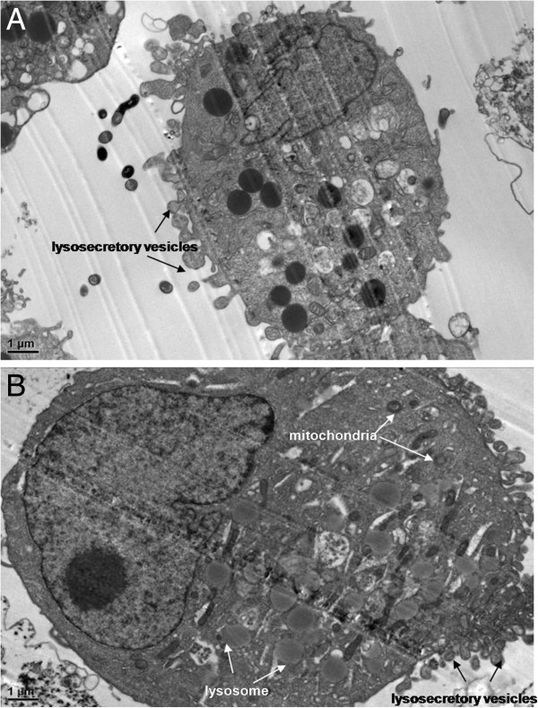
Figure 6.

TEM images of N9 cells treated with SWNHs. (A) N9 cells treated with SWNHs untreated with LPS as control (×15,000 magnification). Scale bar represents 1 μm. (B) N9 cells cultured onto SWNHs-coated dishes (0.85 μg/cm2) for 48 h pre-treated with LPS (×15,000 magnification). Scale bar represents 1 μm. The size of N9 cells pre-treated with LPS and their nucleus were larger than that of control. The apoptotic bodies were observed in cytoplasm. The size of lysosome and mitochondria in N9 cells pre-treated with LPS (B) were larger than that of control (A). A lot of secretory vesicles could be observed outside of cells treated with SWNHs. All data are represented as mean ± SEM.
