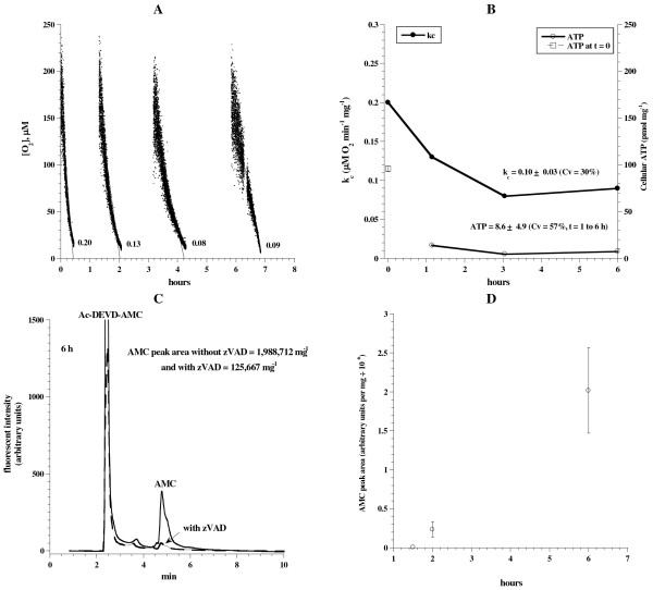
Figure 4.
Liver tissue respiration, ATP content and caspase activity in unoxygenated KH buffer. Liver specimens from a C57Bl/6 mouse were incubated at 37°C in 50 mL KH buffer for up to ~6 h. At indicated time periods, samples were removed from the incubation medium and processed for measurements of O2 consumption, ATP content and caspase activity as described in Methods. Panel A: Representative runs of cellular mitochondrial O2 consumption are shown; t = 0 corresponds to animal sacrifice. The rate of respiration (k, μM O2 min-1) was the negative of the slope of [O2] vs. t. PanelB: The values of kc and ATP are plotted as a function of incubation time. Panel C: HPLC runs of caspase activity at 6 h with and without the pancaspase inhibitor zVAD-fmk. Panel D: AMC peak areas (retention time, ~4.8 min) are shown as a function of incubation time.

