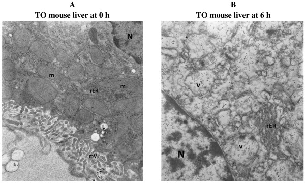
Figure 5.

Electron microscopy images (unoxygenated KH buffer). (A) Liver at time 0 demonstrating hepatocytes with preserved architecture. Note hepatocyte nucleus (N), numerous intact mitochondria (m), rough endoplasmic reticulum (rER), microvilli (mV) and cytoplasmic lipid vacuoles (L). (B) Liver at 6 h demonstrating hepatocytes with numerous cytoplasmic vesicles (v), rough endoplasmic reticulum (rER) and a nucleus (N). Some of the vesicles probably represent swollen disintegrating mitochondria under experimental conditions. Magnification 140,000.
