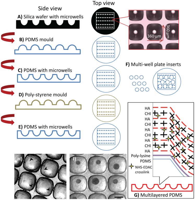
Figure 1. Fabrication of the microwell surface from PDMS replica moulding and surface modification.
A silica wafer having an array pattern of microwells was formed via deep reactive ion etching. The dimensions of the microwells on silica wafer were 360×360×180 (depth) µm (A). This surface was used to cast PDMS, generating a negative surface. PDMS mould having an inverted microwell pattern (B). This surface was then coated in 5% pluronic acid solution, which functioned as a release agent. The coated surface was used to cast a 2 mm thick PDMS sheet having a microarray pattern identical to the original silica wafer (C). Because PDMS-PDMS casting was not reproducible, the PDMS sheet with the microwells was cast with a polystyrene sheet to obtain a plastic mould (D). Using polystyrene mould PDMS sheets with microwells were produced (E). A punch was used to create 2 cm2 discs which fit snuggly into the bottom of a 24-well plate (F). Individual microwell inserts were subsequently surface modified using a CHI/HA electrostatic multilayer; see text for details (G). The chondrocytes spreading on non-modified PDMS microwell surface (cell layers marked with arrowheads) (H). Robust micropellet formation on CHI/HA multilayered PDMS surface (I). Scale bar: 200 µm.

