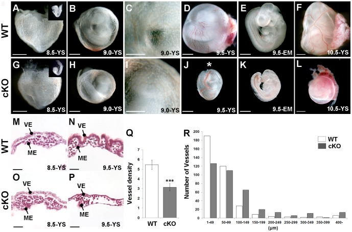
Figure 1. FoxA3-Cre mediated Yy1 cKO deletion results in prominent yolk sac defects at 9.5 dpc.
A-L) Bright field images of WT (A–F) and cKO (G–L) embryos (EM) alone or embryos within their yolk sacs (YS) at the indicated stages. A, G) 8.5 dpc mutant embryos (inset in G) are sometimes slightly delayed compared with WT (inset in A) but display no noticeable yolk sac defects. B–C, H–I) 9.0 dpc mutants display relatively normal yolk sac blood vessel development. D–E, J–K) 9.5 dpc mutant yolk sacs have dilated vessels (asterisk in J) and poor vessel organization (compare D to J). cKO embryos (K) are smaller than WT embryos (E) from the same litter. F, L) While prominent large blood vessels are easily detected in 10.5 dpc WT yolk sacs (F), the yolk sacs of mutants are uniformly pale (L). M–P) A comparison of WT and mutant H&E stained yolk sac sections demonstrates that while no differences are found at 8.5 dpc (M, O), the 9.5 dpc cKO yolk sac (P) has fewer and larger vessels compared with WT (N). Q) Investigation of the same sized area at 9.5 dpc revealed significantly fewer vessels in mutant compared with WT yolk sacs (*** = p<0.001; error bar = standard error). R) A size distribution chart at 9.5 dpc reveals that mutants contain fewer of the small vessels (<100 µm) and more of the larger vessels (>100 µm) compared with WT yolk sacs.

