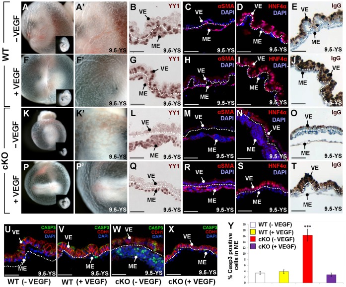
Figure 6. Exogenous VEGF rescues Yy1 cKO yolk sac defects.
A–Y) WT and cKO embryos cultured from 8.5–9.5 dpc in the presence (+VEGF) or absence (−VEGF) of VEGF. A–E) WT cultured embryos display normal yolk sac vasculature (A–A’), typical embryonic size (inset in A) and the presence of YY1 (brown) in the visceral endoderm (VE) and in yolk sac mesoderm (ME; B). In WT yolk sac sections, αSMA (red) surrounds mature vessels (C), HNF4α (red) is expressed in the visceral endoderm (D) and IgG is localized to the apical visceral endoderm (E). F–J) WT embryos cultured with exogenous VEGF display robust yolk sac vasculature (F, F’), YY1 expression in both YS layers (G), normal αSMA in mature vessels (H), typical HNF4α in the visceral endoderm (I) and high levels of apical IgG (J). K–O) Cultured cKO embryos demonstrate poor vascular development, including pooled blood in the proximal yolk sac (K–K’), no YY1 in the visceral endoderm (L), reduced αSMA (M), reduced HNF4α (N) and decreased apical IgG (O) when compared with WT cultured embryos (A–E). P–T) cKO embryos cultured with exogenous VEGF display normal yolk sac vasculature (P–P’) and increased embryo size (inset in P) when compared to cKO embryos cultured without exogenous VEGF (K–K’). cKO embryos cultured with VEGF lack visceral endoderm YY1 (Q) but have increased αSMA in the yolk sac mesoderm (R) and increased levels of HNF4α (S) and apical IgG (T) in the visceral endoderm when compared to untreated cKO embryos (M–O). U–Y) Immunofluorescence against cleaved Caspase-3 (CASP3, green) and CDH1 (red) of sectioned yolk sacs revealed that typical CDH1 expression found in WT (U) and WT cultured with VEGF (V), was downregulated in cultured cKO embryos but more normal visceral endoderm expression restored when cKO embryos were cultured with VEGF (X). Y) Quantification of cleaved Caspase-3 positive cells demonstrates that the addition of VEGF to cKO embryos restores WT levels of apoptosis. *** = p<0.001; error bars = standard error; dotted line is drawn between the visceral endoderm (VE) and mesoderm derivatives (ME) on yolk sac sections.

