
Figure 1.
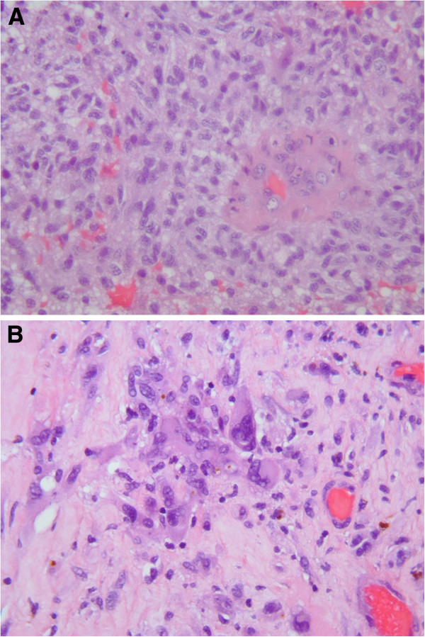
Histopathological evaluation. Hematoxylin and eosin (HE) histologic analysis revealed a highly cellular tumour tissue composed of pleomorphic astroglial cells with hyperchromatic nuclei, mitosis and glomeruloid vascular proliferation, which are a classic histological features in glioblastoma multiforme (A, high-power photomicrograph, original magnification, ×400). After 48Gy chemoradiotherapy, previously treated glioblastoma shows heterogeneos composition with area of coagulative necrosis and hyalinized blood vesels. Nuclear pleomorphism of tumour cells without mitosis were noted (B, high-power photomicrograph, original magnification, ×400).