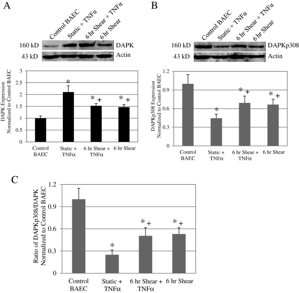
Figure 2.
Shear stress reduced DAPK expression following TNFα treatment on membrane substrates. Western blot analysis for total DAPK expression (A) as well as phosphorylated serine 308DAPK (B) in Control BAEC, Static + TNFα, and 6 hr Shear + TNFα, and 6 hr Shear cells on membrane substrate. Each western blot image is representative of the DAPK or phospho-DAPK protein expression in each particular sample (Top panels). Loading control was assessed with anti-actin antibody on the same blot. We observed an increase total DAPK expression and a decrease of phospho-serine 308 DAPK after TNFα induction alone and in cells that were sheared for 6 h. Relative band intensity was quantified and analyzed based on results from 3 independent experiments, and presented as fold increase over control BAEC for overall DAPK and phospho-serine 308 DAPK (Bottom panels). Expression of total DAPK increased while phospho-serine 308 DAPK decreased following TNFα treatment compared to control BAEC. Adding shear stress to TNFα treatment or shearing cells alone significantly decreased DAPK expression compared to TNFα alone, but the difference was more significant in the phospho-DAPK analysis. Phosphorylated DAPK decreased to close to 50% with the addition of 6 h of shear stress, with or without prior TNFα treatment as compared to a near 26% decrease from TNFα treatment alone. (C) Phosphorylated DAPK as a fraction of total DAPK expression was calculated for each sample group based Western blots. All data represent average ± standard error (n = 3). * P < 0.01 compared to control cells, and + P < 0.05 compared to static TNFα treated cells.

