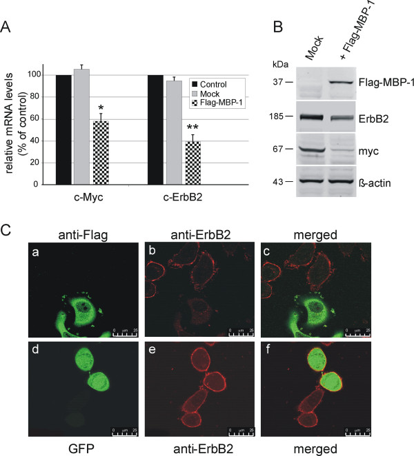
Figure 1.
MBP-1 negatively regulates ERBB2 and c-MYC expression in SKBr3 breast cancer cells. (A) Quantitative analysis of endogenous c-MYC and ERBB2 transcripts by qRT–PCR. SKBr3 cells were transfected with either a vector expressing MBP-1 (pFlag-MBP-1) or an empty vector (mock) and analyzed 48 hrs after transfection. Histograms show fold changes in the expression of c-MYC and c-ERBB2 mRNA after normalization with TBP. Each data point is the average of at least three independent transfection experiments, bars represent standard deviation and p values (* P< 0.05, ** P<0.005) indicate statistical significance. (B) Western blot analysis of myc and ErbB2 proteins in SKBr3 cells overexpressing Flag-MBP-1 and in the mock control. Both mRNA and protein levels were reduced in transfected cells. (C) Representative confocal microscopy images showing the intracellular localization of endogenous ErbB2 protein and either ectopically-expressed Flag-MBP-1 or GFP protein. After transfection, SkBr3 cells expressing Flag-MBP-1 were double-stained with mouse anti-ErbB2 and rabbit anti-Flag antibodies (panels a-c), GFP-expressing cells were single-stained with anti-ErbB2 primary antibodies (panels d-f). Right panels show the merged image of the middle and left panels. Scale bar, 25 um. For supplementary images, see Additional file 2.

