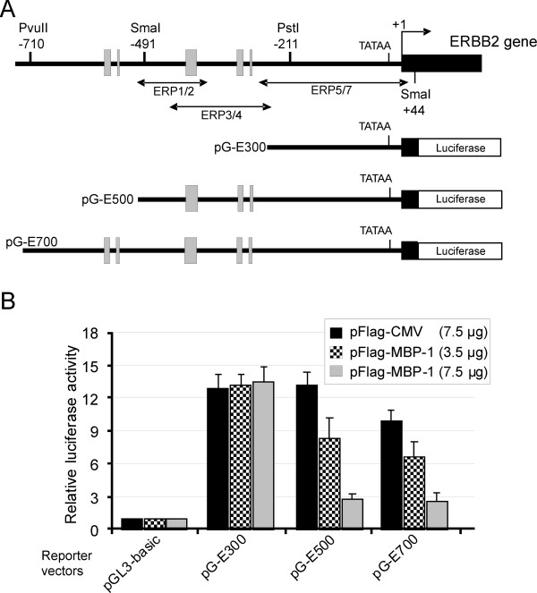
Figure 2.
MBP-1 represses ERBB2 promoter activity. (A) Schematic representation of ERBB2 exon-1 (black box) and 5′-flanking region. The TATA-box, the major transcriptional start site (+1), the position of relevant restriction sites and the location of A/T-rich sequences (gray boxes) are indicated. The numbers refer to the major transcription start site according to NCBI Ref Seq NG_007503.1. Sequences amplified by the three primer sets used in ChiP-qPCR assays are underlined. The schematic structures of the reporter plasmids, containing fragments of the human ERBB2 promoter upstream of the firefly luciferase gene, are shown below (see Additional file 1: Table S1 for details). (B) Functional analysis of the ERBB2 promoter in SkBr3 cells. Cells were transiently cotransfected with each reporter plasmid and two different amounts of the vector expressing Flag-MBP-1 (3.5 or 7.5 μg) or with the highest amount of the empty vector pFlag-CMV (7.5 ug). Values of luciferase activity, corrected for transfection efficiency, are expressed relative to the activity obtained with the pGL3-basic plasmid to which was assigned the value of 1. Each data point is the average of at least three independent experiments and the error bars represent SD.

