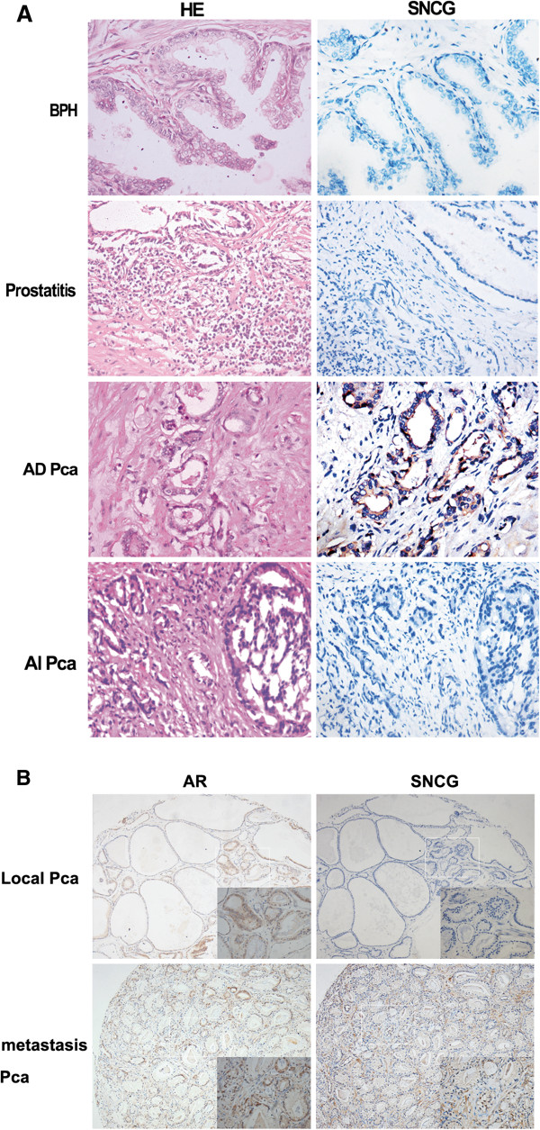
Figure 6.

SNCG protein expression is associated with human prostate cancer progression and metastasis. (A) SNCG protein expression detected by immunohistochemical (IHC) staining was representative in a series of human prostate tissues on a tissue microarray (TMA). Benign prostatic hyperplasia tissues and prostatitis showed no SNCG expression in either epithelial or stromal regions. Strong staining of SNCG protein displayed in androgen-dependent prostate cancers and negative SNCG staining presented in androgen-independent prostate cancers. The left panel shows H&E staining and the right panel shows SNCG IHC staining (400×). (B) Representative immunohistochemical staining of SNCG (right line) or AR (light line) protein in local or metastatic human prostate cancer tissues.
