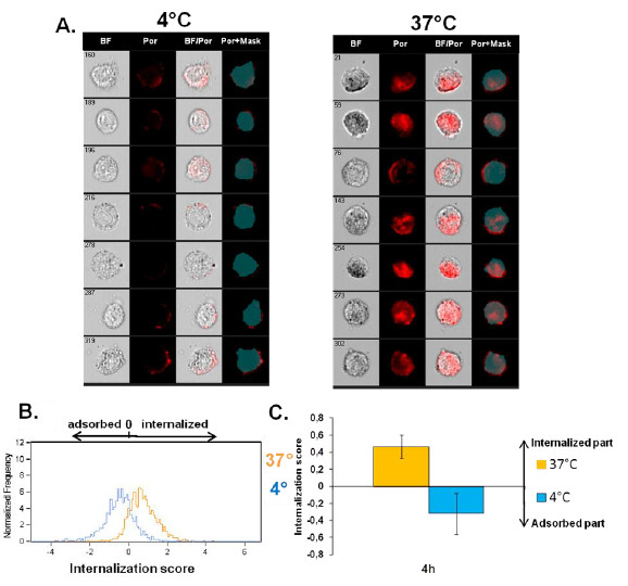
Figure 5.
Determination of 100 nm-Por-SiO2 uptake by NCI-H292 cells by imaging flow cytometry. A. Representative images captured by the Amnis ImageStreamX Flow Cytometer of cells treated with 100 nm-Por-SiO2 for 4 h at 37°C or 4°C. First column shows brightfield (BF) images of the cells, second column shows images of fluorescence of porphyrine (Por), third column shows fluorescence merged with the brightfield images of the cells (BF/Por) and forth column shows the applied mask eroded for 3 μm and porphyrine fluorescence (Por+Mask). B. and C. Internalization score (IS) calculated by Amnis IDEAS software: distribution of IS of at least 500 cells treated for 4 h at 37 °C or 4°C (B.), and corresponding mean value of IS ± SD of six independent experiments (C.).

