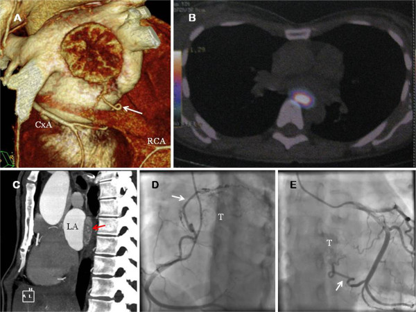
Figure 1.

A. Computed tomography imaging reconstruction of the heart. The tumor is showed. The collateral vessel (white arrow) from the circumflex artery (CxA) is observed. B. 18-F DOPA PET showed a 16 × 25 × 36 mm extracardiac mass protruding within the LA. Presence of metabolic activity was detected. C. Computed tomography (axial view) showed the presence of the large mass (red arrow). D. Coronary angiography. The coronary blood supply of the superior aspect of the paraganglioma (T) emerged from a large collateral vessel (white arrow) from the right coronary artery (RCA). E. Coronary angiography. Proximal and distal collaterals from CxA were the feeding vessels (white arrow) of the inferior aspect of the tumor (T).
