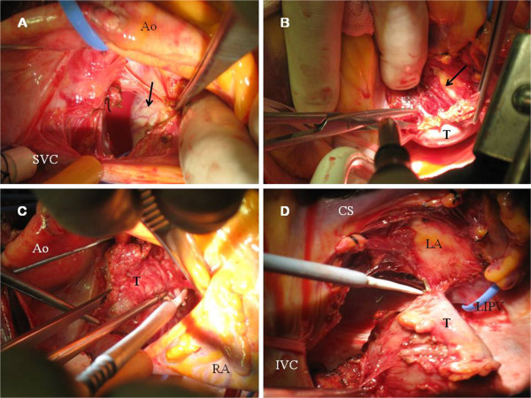
Figure 3.

A. Surgeon’s view. Superior aspect of the tumor (black arrow) is showed. Neovascularization of the smooth surface is observed. B. Endocardium of the LA was exposed (black arrow) during surgical excision (suspended heart). C. Surgeon’s view. Dissection of the superior aspect of the tumor (T). D. Paraganglioma (T) after totally excision from the LA. IVC: Inferior vein cava. LIPV: Left inferior vein cava.
