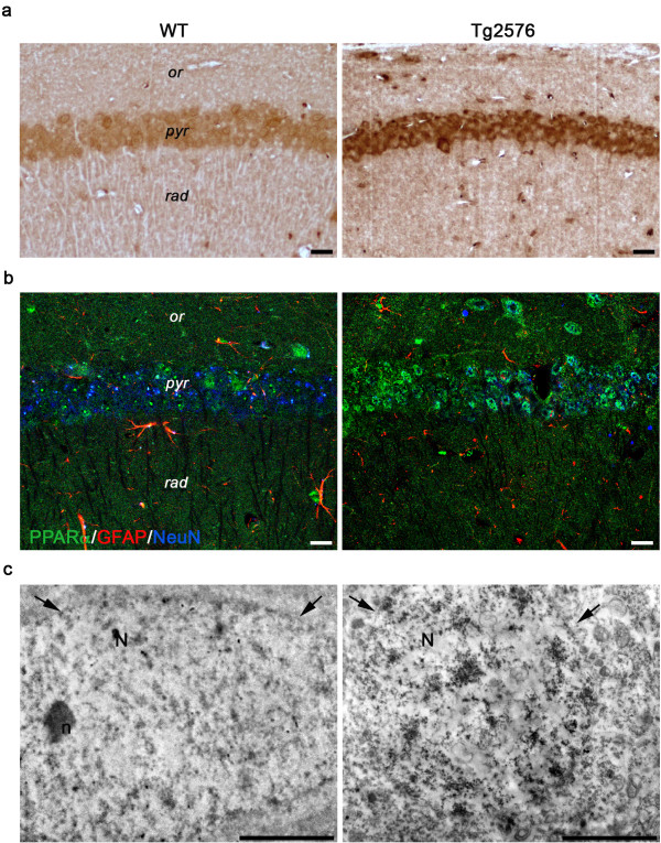
Figure 9.
PPARα immunolocalization in 3-month-old WT and Tg mice. (a) PPARα immunohistochemistry in CA1 pyramidal cell layer shows a higher immunoreactivity in Tg hippocampus, than in its WT counterpart. or, stratum oriens; pyr, stratum pyramidale; rad, stratum radiatum. Scale bars, 25 μm. (b) Confocal image of triple immunofluorescence for PPARα (green) in combination with GFAP (red) and NeuN (blue) demonstrates the presence of PPARα-positive neurons and astrocytes. Note the brightness of green signal in the Tg section. Scale bars, 25 μm. (c) PPARα pre-embedding immunoelectron microscopy of CA1 pyramidal neurons shows the nuclear localization of the immunoreaction product, especially concentrated in the Tg nucleus. Arrows indicate the nuclear envelope. N, nucleus; n, nucleolus. Scale bars, 2 μm.

