
Figure 15.
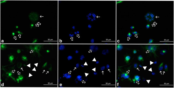
a-f Organization of vimentin in A549 cells after treatment with etoposide - fluorescence microscopic examination (vimentin indirect labeling, secondary antibody was goat anti-mouse IgG-BODIPY). a-c 0.75 μM etoposide, d-f 3 μM etoposide; a, d vimentin labeling, b, e DAPI, c, f merged. Selected photographs presenting rare nuclei with chromatin structures bearing resemblance to senescence-associated heterochromatin foci (thin arrows). These cells featured either disappearing (a, c) or more regularly woven (d, f) vimentin scaffold, devoid of threads/foci typical of nuclear invaginations and/or multinucleated cells. Thick arrows point at septum-like structures between sister nuclei (a, c), vimentin aggregates in the perinuclear area of multinucleated cells/cells possessing abnormally-shaped nuclei (d, f) or nuclei of these cells (b, e). Double thick arrows - putative budding-like structures (a, c); arrowheads - diffuse vimentin staining around nuclei with features of karyorrhexis or karyolysis (d, f). Results are indicative of five independent experiments. Bar 50 μm.