
Figure 4. Dehydratase Active Site Arrangement A.
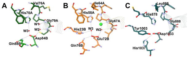
Potential hydrogen bonds are shown with grey lines, red spheres represent resolved water molecules, and residues are shown as sticks and colored by element. The beginnings of the hot dog helices are shown with faint line ribbons. The images are drawn to scale and superimposable. (A) The active site of E. coli FabA (PDB#1MKB), an example of an SHD DH, in green. (B) The active site of H. pylori FabZ (PDB#2GLL), another SHD DH, in orange. The green sphere represents a chloride ion. (C) The active site of S. scrofa (porcine) FAS DH domain (PDB#2VZ8), a DHD DH, in dark cyan. The crystal structure for this enzyme did not include resolved water molecules.