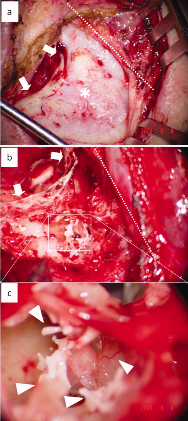
Fig. 3.

Regenerated mastoid air cells after reopening the mastoid cavity at the second-stage operation (left ear) (white dotted line, temporal line; white asterisk, regenerated mastoid cortex bone; white arrow, posterior wall of external auditory meatus; white arrow head, artificial pneumatic bones). (a.) The mastoid cortex bone showed complete regeneration 1 year after the first-stage operation. (b.) Regenerated mastoid air cells and good aeration were observed after reopening the mastoid cavity. (c.) Enlargement of regenerated mastoid air cells. [Color figure can be viewed in the online issue, which is available at wileyonlinelibrary.com.]
