Figure 3.

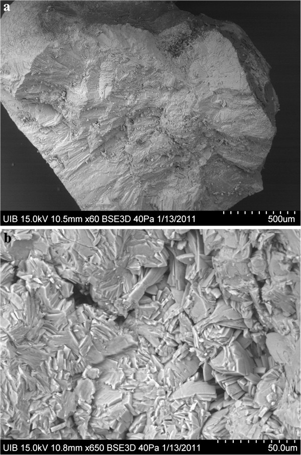
Scanning electron microscopy images of a section of a COM papillary calculus from patient 3. (a) General view of the calculus section. (b) Calculus core formed by the intergrowth of COM crystals and organic matter.

Scanning electron microscopy images of a section of a COM papillary calculus from patient 3. (a) General view of the calculus section. (b) Calculus core formed by the intergrowth of COM crystals and organic matter.