
Figure 2. Characteristics of fibrocytes in tissues and in the circulation.
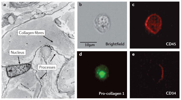
a | A representative electron micrograph of a CD34+ fibrocyte in the dermis of a patient with nephrogenic sclerosing fibrosis. The presence of a large nucleus, collagen fibres and extensive processes are indicated by the arrows. In three dimensions, this cell is spindle shaped. Image is reproduced, with permission, from REF. 91 © (2001) Lippincott Williams & Wilkins. b | A brightfield image of a fibrocyte obtained from the circulation of a normal human. c,d,e | Confocal images of this fibrocyte following staining for CD45 (bright red), intracellular pro-collagen I (green) and CD34 (dark red).