
Figure 3.
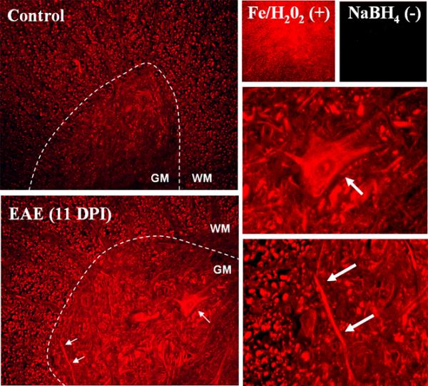
Immunohistochemical localization of protein carbonyls in the rat spinal cord of EAE animals. Carbonyls in thoracic spinal cord sections of a control and an EAE rat (score = 3.0) were detected by immunohistochemistry as described in Materials and Methods. Images show carbonyls the spinal cord ventral horn. Both neuronal cell bodies and axons are heavily stained in the EAE sample (arrows and insets). The carbonyls increase is seen throughout the spinal cord tissue, not localized around lesions. Tissue sections were treated with Fe/H2O2 to generate carbonyls (positive control) or NaBH4 to block endogenous carbonyls (negative control).