Fig.4.
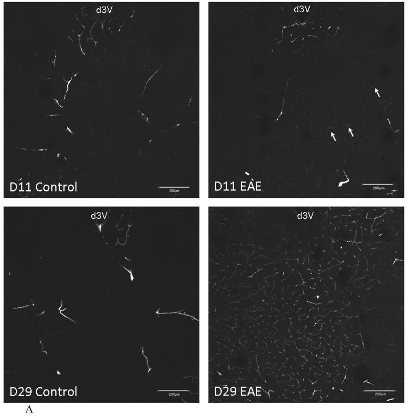
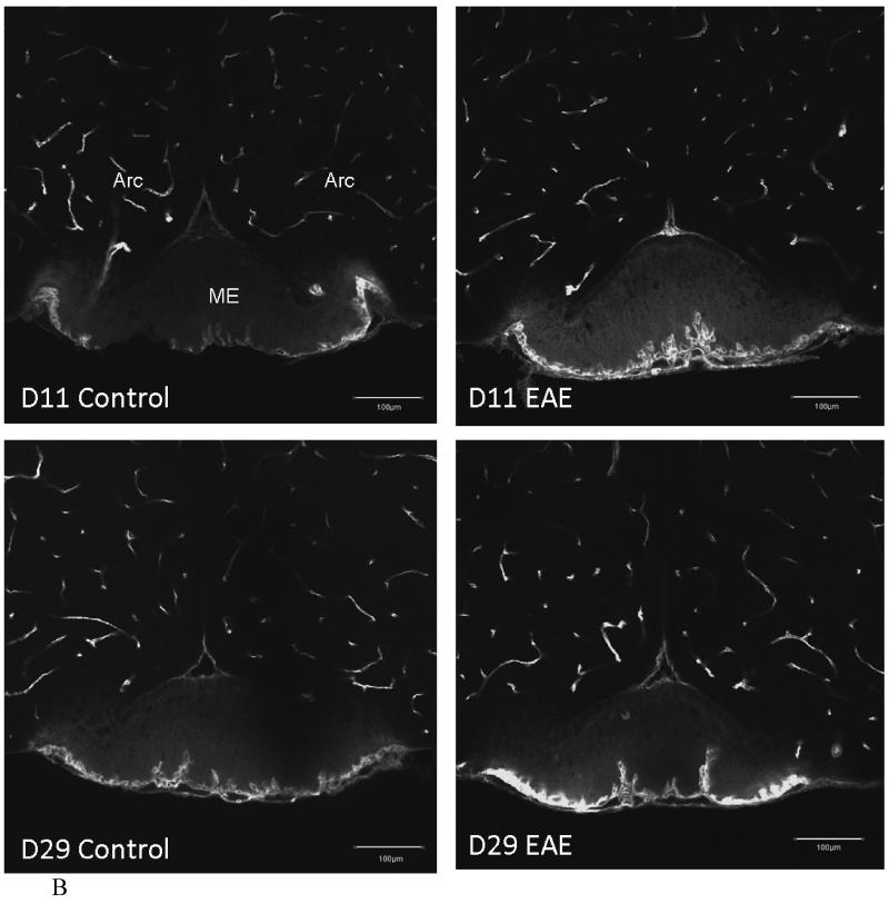
Vascular space of control and EAE mice after induction with PLP and CFA.
(A) In the striatum and thalamus, the vascular space was slightly increased in the EAE mouse 11 days after PLP treatment (d11) in the area under the dorsal 3rd ventricle (d3V) (arrows), and was significantly increased at d29, compared with the controls.
(B) The blood vessels beneath the median eminence (ME) also showed a similar pattern in EAE mice. There is no significant difference between control and EAE mice in the vascular space in most of the brain regions, as shown in the arcuate nucleus (Arc). There was no BBB leakage seen in EAE mice within this perfusion time frame but the diffusion of FITC-albumin can be seen in CVOs, such as the ME (scale bar = 200 μm).
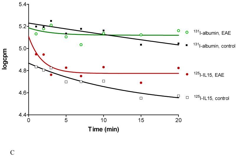
Fig.4C. Disappearance of 125I-IL15 and 131I-albumin from blood after iv bolus injection. EAE was associated with altered kinetics and a shortened half-life for both substances.


