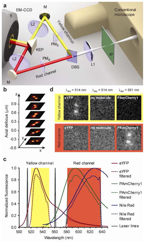
Figure 1.
Multicolor imaging with DH-PSF phase masks. a Schematic of the multicolor DH-PSF microscope. The conventional wide-field inverted fluorescence microscope is at the right with the conventional image plane IP. The dichroic mirror DM splits the emission into two spectrally distinct optical paths for proper phase modulation by two different phase masks PMa and PMb. Lenses L1 and L2 in each path form a 4f optical processing system, with the phase masks located one focal length away from the first lens L1 and from the second lens L2. The knife edge prism KEP directs both paths onto two adjacent regions of the same EMCCD. b For a single emitter, the DH-PSF microscope generates two focused spots on the detector, which revolve around each other as a function of z position of the emitter. c Raw and filtered emission spectra of fluorescent labels used in this work. The various filters in the optical system create two spectrally distinct detection channels. d Even bright localizations (~4000 detected photons) of eYFP and PAmCherry1 do not produce a detectable signal in the spatially corresponding region of the opposing channel. Also shown are the background levels for 514 nm excitation, when no molecules are present (middle column).

