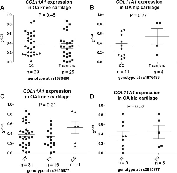
Figure 2.
Columnar scatter plots of the quantitative expression of COL11A1 in OA cartilage cDNA stratified by A) genotype at the LDH associated SNP rs1676486 in knee cases, B) genotype at the LDH associated SNP rs1676486 in hip cases, C) genotype at the OA associated SNP rs2615977 in knee cases, and D) genotype at the OA associated SNP rs2615977 in hip cases. Due to the low frequency of TT homozygotes at rs1676486 the analysis in A) and B) was between CC homozygotes and T-allele carriers (CT and TT combined) at this SNP. Due to the absence in the hip strata of GG homozygotes at rs2615977 the analysis in D) was between TT homozygotes and TG heterozygotes at this SNP. n is the number of patients studied for each group. The horizontal lines in each plot represent the mean and the standard error of the mean. P-values were calculated using a Mann–Whitney U test for A), B) and D) and a Kruskal-Wallis test for C).

