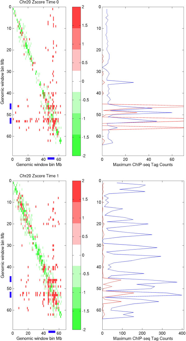
Figure 1.
Chromosomal interaction hotspots. Upper panel: intra-chromosomal interaction for chromosome 20 at control condition; down panel: intra-chromosomal interaction for chromosome 20 at E2-treated condition; right panel, red smooth line represents detected number of ERα binding sites in the region (1 Mb resolution), and blue smooth line is the maximum read counts in the region (1 Mb resolution); left panel, chromosomal interaction hot regions (within amplified regions) that identified by this study are colored by blue bar at X-axis and Y-axis, respectively; positive and negative Z-scores are colored by red and green color, which indicate the observed chromosome region has higher and lower interaction frequency than the average, respectively.

