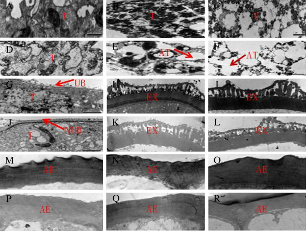
Figure 2.

TEM analysis of anthers in the WT and GMS mutant. Transverse sections of the (A-C) WT and (D-F) GMS mutant tapetum at the (A, D) meiosis, (B, E) tetrad, and (C, F) uninucleate microspore stages. Extine development in (G-I) WT and (J-L) GMS mutant microspores at the (G, J) meiosis, (H, K) tetrad, and (I, L) uninucleate microspore stages. Outer wall of anther epidermal cells in the (M-O) WT and (P-R) GMS mutant at the (M, P) meiosis, (N, Q) tetrad, and (O, R) uninucleate microspore stages. T: Tapetal layer; AT: abnormal tapetal layer; UB: Ubisch body; NUB: no Ubisch body; Ex: extine; AE: anther epidermal cuticle. Bars = 2 μm (A-L), 1 μm (M-R).
