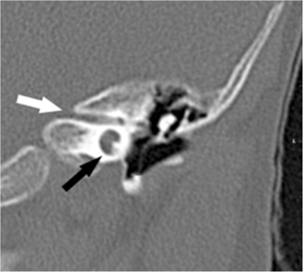
Figure 1.

High resolution CT of the left petrosus bone. The coronal view demonstrates the widened single basal cochlear turn (black arrow) and the narrowing of the auditory canal (white arrow). The patient did not show an enlarged vestibular aqueduct (EVA) but similar findings of the right petrosus bone.
