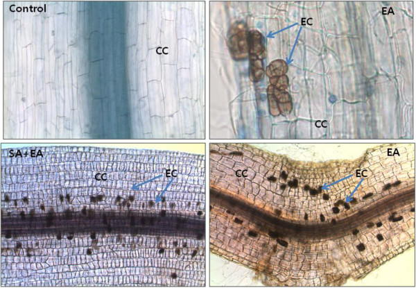
Figure 4.
Light micrographs of endophyte P. resedanum – associated with host plant’s root. (Control) shows the light microscopic image of endophyte-free control plants (two weeks old). Bar = 200 μm. (EA) pepper root infected with P. resedanum after one week of inoculation. Microsclerotiums in the form of yeast-like cells were extensively colonized in the middle and inner cortex of the root. Bar = 40 μm; Bar = 100 μm. (SA+EA) presence of brownish yeast-like cells pericycle regions of the roots. Bar = 100 μm. The root samples were stain with tryptophan blue (0.8%). In the micrographs, CC = cortex cells; EC = endophyte cell.

