Figure 3.
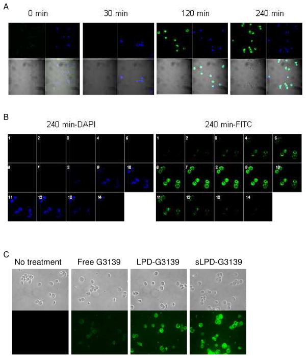
Internalization of sLPD-G3139 in KB cells
The cells were incubated with 500 nM sLPD-G3139 (containing 10% fluorescent FAM-G3139) at 37 °C. At different time points, unbound sLPDs were removed by washing with PBS and cellular nuclei were stained by DAPI. The cell were then mounted to the slide and observed under a fluorescence microscope. Blue color indicated nuclei stained by DAPI and green color indicated FAM-labeled G3139.
Panel A. Cells were treated with sLPD-G3139 spiked with 10% FAM-G3139 (green) at 37°C for 30, 120 and 240 min., respectively, stained by DAPI (blue) and visualized on a confocal microscope.
Panel B. Optical sections (14 total) of cells were collected after 4 hr by incremental scanning along the z-axis at a spacing of 0.45 μm.
Panel C. Cells were treated with sLPD-G3139 and visualized on a fluorescence microscope.

