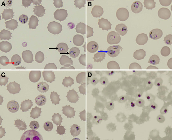
Figure 1.
Blood smear before treatment (A-C) and 1 day post-treatment (D). Arrow in red shows parasite with distantly parted chromatin dots, whereas arrow in black shows parasite with chromatin dot located within the “ring” form (A). Multiple-infection was found (B). Compacted “band” form trophozoite can be seen here (pointed by blue arrow). P. knowlesi in highly amoeboid forms are shown in (C). The characteristic golden-brownish pigments can be seen within the parasites.

