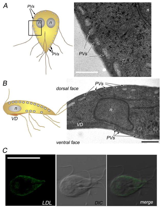
Fig. 1. Endosomal-lysosomal peripheral vacuoles in Giardia lamblia.
(A) On the left, cartoon of a Giardia trophozoite. The oval-shape peripheral vacuoles (PVs) are illustrated beneath the plasma membrane. The region analyzed by transmission electron microscopy (TEM) is outlined. On the right, transmission electron microscopy of the region corresponding to the median section, parallel to the dorsoventral axis of the cell, showing the PVs distributed underneath the plasma membrane. n: nucleus. Bar, 500 nm. (B) On the left, cartoon of a trophozoite viewed laterally through the ventral groove. The region showing the array of the PVs analyzed by TEM is bordered. On the right, transmission electron microscopy of a transverse section, showing the PVs distributed below the dorsal and ventral sides of the plasma membrane. n: nucleus. VD: ventral disc (attachment organelle). Bar, 500 nm. (C) BODIPY-LDL fluorescence and confocal microscopy show the distribution of LDL in the PVs underneath the plasma membrane (Rivero et al. 2010). DIC: differential interference contrast microscopy. Bar, 10 μm.

