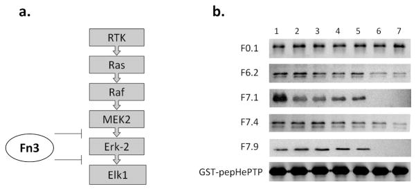
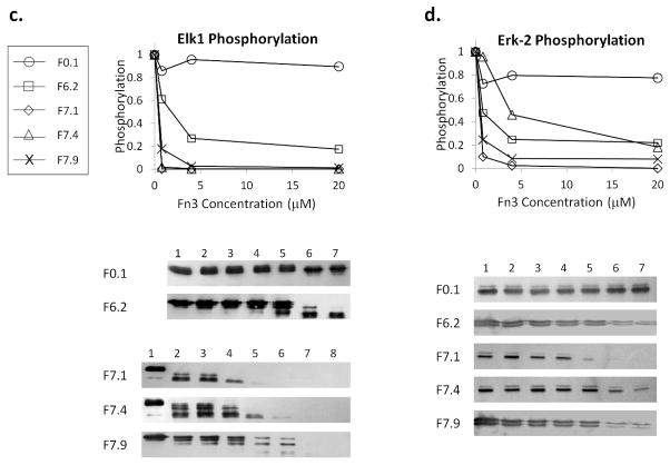
Figure 3. Functional assays of engineered Fn3.

a. The components of mammalian ERK pathway. The binding of the MEK2 D-peptide mediates Erk-2 phosphorylation, and the binding of the Elk1 D-peptide mediates Elk1 phosphorylation. The engineered monobodies should disrupt both interactions.
b. GST-pepHePTP was used to pull down Erk-2 in the presence of Fn3 competitors. The precipitated Erk-2 was visualized by Western blot using M2. F0.1 is a control monobody that binds MBP (22). Competitor concentrations in Lanes 1 – 7: 0, 0.016, 0.08, 0.4, 2, 10, and 50 μM.
c. In vitro phosphorylation of Elk1(307-428) by Erk-2 in the presence of Fn3 competitors was analyzed by Western blot. Competitor concentrations: (For F0.1 and F6.2) 0, 0.0064, 0.032, 0.16, 0.8, 4, and 20 μM. (For F7.1, F7.4, and F7.9) 0, 0.0013, 0.0064, 0.032, 0.16, 0.8, 4, and 20 μM. The phospho-Elk1 band was quantified using ImageJ and normalized to Lane 1. Only the intensities corresponding to three highest Fn3 concentrations and Lane 1 are plotted for clarity.
d. In vitro phosphorylation of Erk-2 by MEK2 in the presence of Fn3 competitors. Competitor concentrations: 0, 0.0064, 0.032, 0.16, 0.8, 4 and 20 μM. The intensity of the phospho-Erk-2 bands was quantified and normalized to Lane 1. Only select intensities are plotted for clarity.

